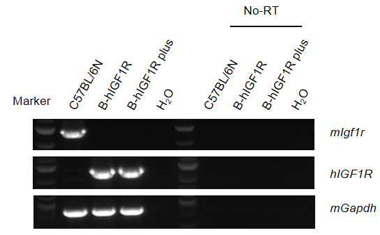
mRNA Expression Analysis of human IGF1R in B-hIGF1R Mice Plus
Strain-specific analysis of IGF1R mRNA expression was performed in wild-type C57BL/6N mice, B-hIGF1R mice, and B-hIGF1R mice plus by RT-PCR.
Kidney RNA was isolated from wild-type C57BL/6N mice (+/+), homozygous B-hIGF1R mice (H/H), and homozygous B-hIGF1R mice plus (H/H). cDNA libraries were synthesized by reverse transcription followed by PCR with IGF1R primers. Mouse Igf1r mRNA was detectable in wild-type C57BL/6N mice. Human IGF1R mRNA was detectable in homozygous B-hIGF1R mice and homozygous B-hIGF1R mice plus, but not in wild-type mice. No-RT indicates no reverse transcription.
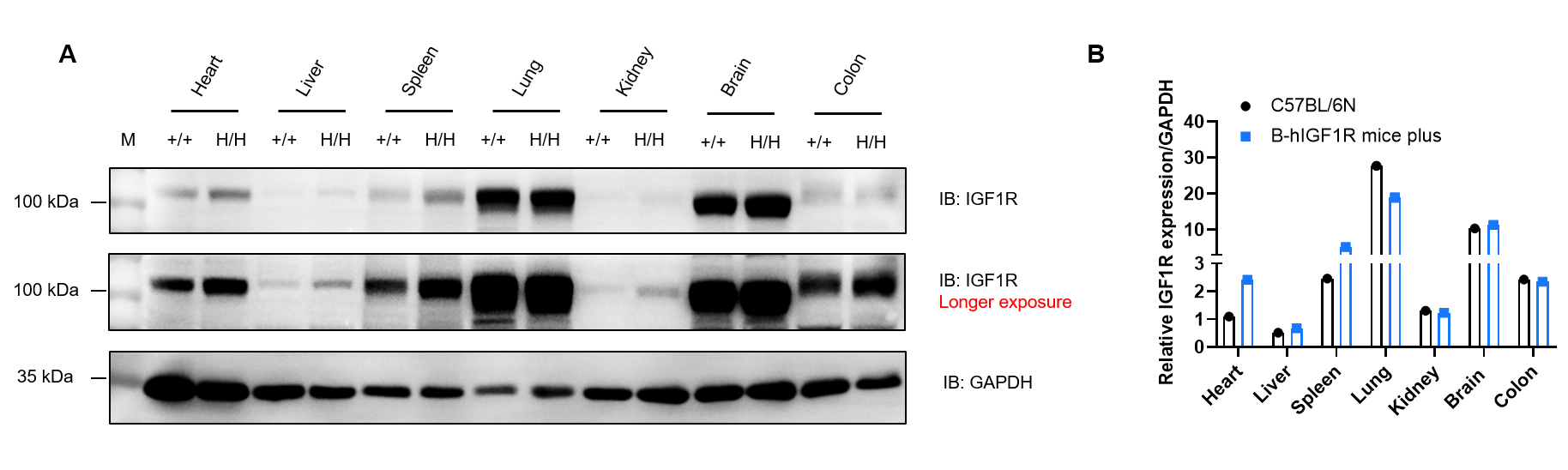
Protein Expression Analysis of IGF1R in B-hIGF1R Mice Plus
Western blot analysis of IGF1R protein expression was performed in wild-type C57BL/6N mice and homozygous B-hIGF1R mice plus.
Heart, liver, spleen, lung, kidney, brain, and colon tissues were collected from wild-type C57BL/6N mice (+/+) and homozygous B-hIGF1R mice plus (H/H) and analyzed by western blot using anti-IGF1R antibody (CST, 3027S). A total of 40 μg protein was loaded for western blot analysis, and GAPDH was used as an internal control. IGF1R was detectable in both C57BL/6N and B-hIGF1R mice plus because the antibody cross-reacts between human and mouse IGF1R. Relative IGF1R protein levels were normalized to GAPDH.
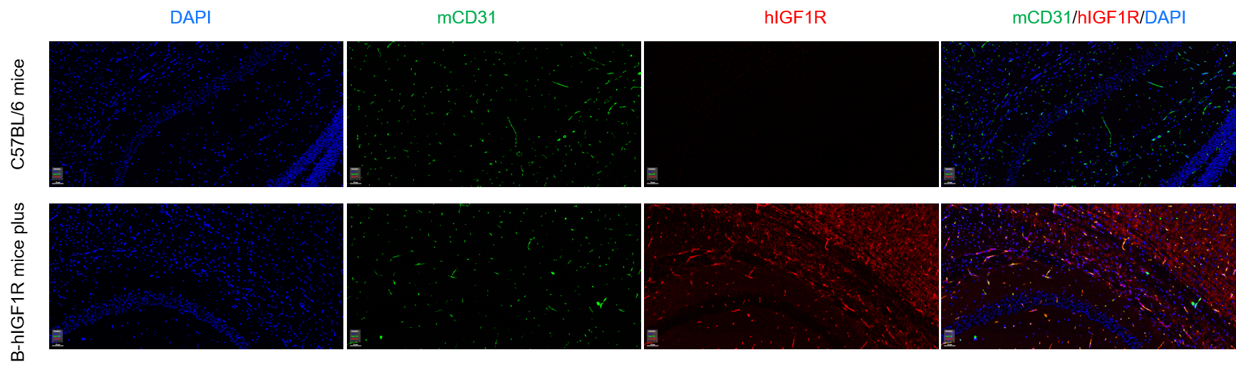
Protein Expression Analysis of IGF1R in Brain Micro-Vessels
Immunofluorescence analysis of IGF1R expression in isolated brain micro-vessels was performed in wild-type C57BL/6 mice and homozygous B-hIGF1R mice plus.
Brain micro-vessels were collected from wild-type C57BL/6 mice (+/+) and homozygous B-hIGF1R mice plus (H/H) and analyzed by immunofluorescence using anti-mCD31 antibody and anti-hIGF1R antibody. Human IGF1R was detectable in brain micro-vessels of homozygous B-hIGF1R mice plus, but not in wild-type mice.
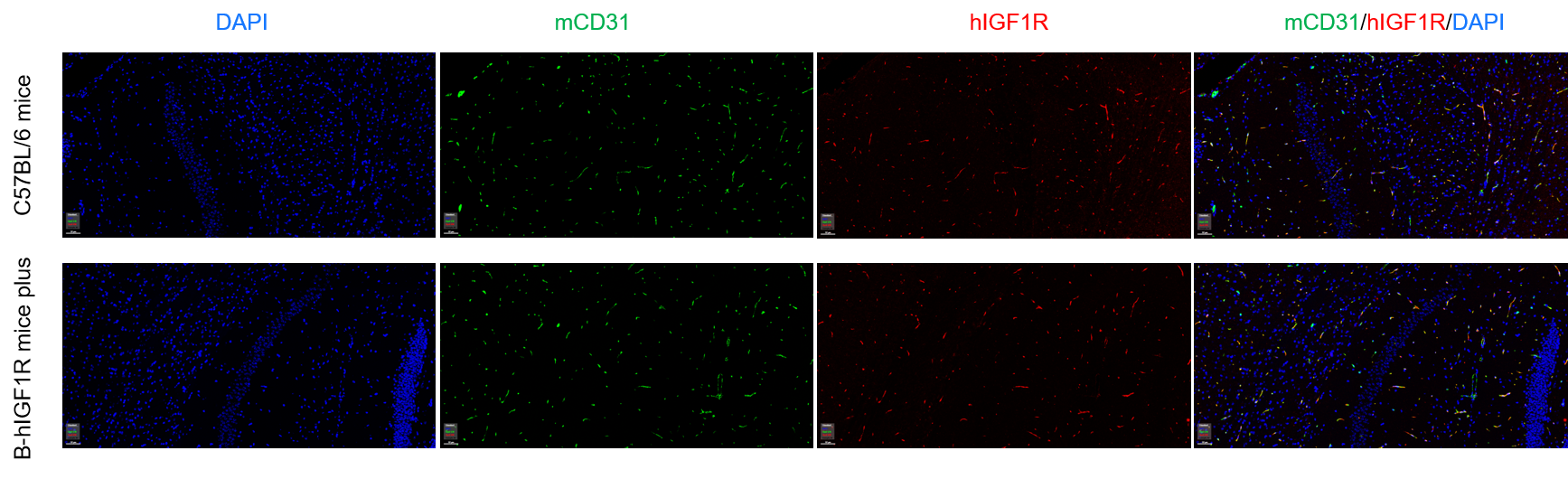
Immunofluorescence analysis of IGF1R expression in isolated brain micro-vessels was performed using an anti-IGF1R antibody that cross-reacts between human and mouse IGF1R.
Brain micro-vessels from wild-type C57BL/6 mice (+/+) and homozygous B-hIGF1R mice plus (H/H) were stained with anti-mCD31 antibody and anti-IGF1R antibody (CST, 3027S). IGF1R signal was detectable in both wild-type C57BL/6 mice and homozygous B-hIGF1R mice plus because the antibody recognizes both human and mouse IGF1R.
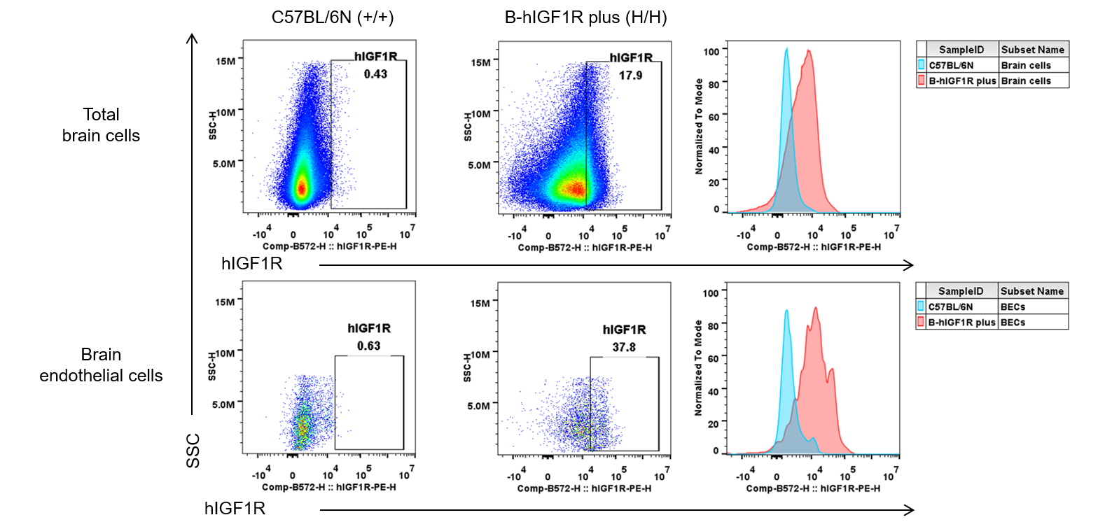
Human IGF1R Expression in Brain Endothelial Cells
Strain-specific IGF1R expression in wild-type C57BL/6N mice and homozygous B-hIGF1R mice plus was analyzed by flow cytometry.
Brain cells were collected from wild-type C57BL/6N mice (+/+) and homozygous B-hIGF1R mice plus (H/H) (male, 7-week-old, n=1), and analyzed with anti-human IGF1R antibody (BioLegend, 351805). Human IGF1R was detectable in total brain cells and brain endothelial cells of homozygous B-hIGF1R mice plus, supporting use of B-hIGF1R mice plus for brain endothelial IGF1R and CNS delivery studies.
Frequency of Leukocyte Subpopulations in B-hIGF1R Mice Plus
Frequency of leukocyte subpopulations in spleen was analyzed by flow cytometry in B-hIGF1R mice plus.
Splenocytes were isolated from wild-type C57BL/6N mice (male, n=3, 7-week-old) and homozygous B-hIGF1R mice plus (male, n=3, 7-week-old). Flow cytometry was used to evalsuate T cells, B cells, NK cells, dendritic cells, neutrophils, monocytes, macrophages, CD4+ T cells, CD8+ T cells, and Tregs. Leukocyte subpopulation frequencies in B-hIGF1R mice plus were similar to wild-type C57BL/6N mice. Blood and lymph node immune cell profiles were also comparable (data not shown). Values are expressed as mean ± SEM. Significance was determined by two-way ANOVA. *p < 0.05, **p < 0.01, ***p < 0.001.
Function Assay of IGF1R in B-hIGF1R Mice Plus
Western blot analysis of phospho-IGF1R/AKT protein expression was performed to evalsuate functional IGF1R signaling in B-hIGF1R mice plus.
Kidney cells from wild-type C57BL/6N mice (+/+), homozygous B-hIGF1R mice (H/H), and homozygous B-hIGF1R mice plus (H/H) were cultured with figitumumab (1 μg/mL) and/or recombinant human IGF1 protein (200 ng/mL). Cells were analyzed by western blot using anti-IGF1R, anti-phospho-IGF1R, and anti-phospho-AKT antibodies. IGF1 induced IGF1R phosphorylation in wild-type and B-hIGF1R mice plus, while figitumumab-analog suppressed IGF1-induced activation of humanized IGF1R in B-hIGF1R mice plus. Phosphorylation of endogenous mouse IGF1R remained unaffected, reflecting the species-related binding profile of figitumumab.
Hematology Analysis of B-hIGF1R Mice Plus
Complete blood count (CBC) was performed to evalsuate hematological characteristics of B-hIGF1R mice plus.
Blood was collected from male and female C57BL/6JNifdc mice and B-hIGF1R mice plus (n=10, 6-week-old) for CBC analysis. No differences were observed in measured hematology parameters between C57BL/6JNifdc mice and B-hIGF1R mice plus, indicating that humanization of IGF1R does not alter blood cell composition or morphology. Values are expressed as mean ± SEM.
Biochemistry Analysis of B-hIGF1R Mice Plus
Blood chemistry tests were performed to evalsuate serum biochemistry in B-hIGF1R mice plus.
Serum from male and female C57BL/6JNifdc mice and B-hIGF1R mice plus (n=10, 6-week-old) was collected for biochemistry analysis. No differences were observed in measured blood chemistry parameters between male C57BL/6JNifdc mice and male B-hIGF1R mice plus, indicating that humanization of IGF1R does not change blood biochemistry. Values are expressed as mean ± SEM.
Growth Curve Analysis of B-hIGF1R Mice Plus
Body weight growth curve analysis was performed in B-hIGF1R mice plus.
Ten male and ten female homozygous B-hIGF1R mice plus were randomly selected, and body weight was collected once per week. The minimum and maximum weights in the table are calculated as average ± SD. The body weight growth curve of B-hIGF1R mice plus was consistent with that of wild-type mice, supporting normal growth and development after IGF1R humanization.
Efficacy Study of Anti-IGF1R Antibody in HFD-Induced B-hIGF1R Mice Plus
Efficacy of anti-IGF1R antibody was evalsuated in high-fat diet (HFD)-induced B-hIGF1R mice plus.
B-hIGF1R mice plus were fed a high-fat diet to induce obesity. Body weight change was monitored after HFD induction and after treatment with anti-IGF1R antibody provided by a client. Anti-IGF1R antibody significantly inhibited HFD-induced body weight gain, as shown by lower body weight and reduced body weight change in the treatment group compared with the HFD-vehicle group. ***p < 0.001. Values are expressed as mean ± SEM.
Note: This experiment is a collaborative validation project with the client.
Metabolic Efficacy Study of Anti-IGF1R Antibody in HFD-Induced B-hIGF1R Mice Plus
Metabolic efficacy of anti-IGF1R antibody was evalsuated in HFD-induced B-hIGF1R mice plus.
B-hIGF1R mice plus were fed a high-fat diet to induce obesity and treated with anti-IGF1R antibody provided by a client. Food intake, blood glucose, intraperitoneal glucose tolerance test (IPGTT, 15% D-glucose), and insulin tolerance test (ITT, 0.5 U/kg) were analyzed after treatment. Anti-IGF1R antibody affected food intake and metabolic response in HFD-induced B-hIGF1R mice plus. Values are expressed as mean ± SEM. Significance was determined by ordinary one-way ANOVA. *p < 0.05, **p < 0.01, ***p < 0.001.
Note: This experiment is a collaborative validation project with the client.
FAQ section
Q1: What are B-hIGF1R mice plus?
B-hIGF1R mice plus are target-humanized mice expressing a chimeric IGF1R protein with human IGF1R extracellular and transmembrane regions, enabling evalsuation of human IGF1R-targeted therapeutics in vivo.
Q2: Why is IGF1R an important therapeutic target?
IGF1R is a receptor tyrosine kinase involved in IGF signaling, cell growth, survival, metabolism, and transformation, making IGF1R relevant to oncology, metabolic disease, obesity, diabetes, and CNS delivery research.
Q3: How was IGF1R expression validated in B-hIGF1R mice plus?
Human IGF1R mRNA was validated by RT-PCR, IGF1R protein was detected by western blot across multiple tissues, and human IGF1R expression was confirmed in brain endothelial cells and brain micro-vessels.
Q4: Can B-hIGF1R mice plus be used for functional antibody studies?
Yes. Figitumumab-analog suppressed IGF1-induced activation of humanized IGF1R in B-hIGF1R mice plus, supporting functional evalsuation of human IGF1R-targeted antibodies.
Q5: What are the main applications of B-hIGF1R mice plus?
Applications include anti-IGF1R antibody efficacy studies, IGF1R pathway pharmacology, obesity and metabolic disease research, oncology studies, brain endothelial IGF1R research, and preclinical safety evalsuation.
* When publishing results obtained using this animal model, please acknowledge the source as follows: The animal model [B-hIGF1R mice plus] (Cat# 111974) was purchased from Biocytogen.